What makes poison dart frog resistant to their on poison?
This article was originally posted at Amazing Zoology. View original article at http://amzoo.in/sksq7
Contents
Poison Dart Frog
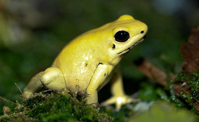
Golden poison frog (Phyllobates terribilis) is one of the deadliest poisonous animals in the world. These animals are endemic to the Pacific coast of Colombia. An average one milligram of poison available in one frog is enough to kill 10-20 humans[1]. Indigenous people carefully expose the frog to the heat of a fire, and the frog exudes small amounts of poisonous fluid. The tips of arrows and darts are soaked in the fluid. Their deadly effect kept for over two years[2]. The interesting question is, what makes the frog itself resistant to this deadly poison!
How is the toxin produced?
These animals store Batrachotoxin, a steroidal alkaloid toxin in their skin glands. This toxin is not actually synthesized by the frogs. When these frogs are removed from their natural habitat and bred in captivity, they lose skin toxicity. This lead to the conclusion that the frogs synthesise the toxin from their diet.
The toxicity of these frogs appear from the consumption of small insects or other arthropods. Recently it has been suggested that the toxin might be derived from a small family of beetles called Melyridae. One species of this beetle is found to produce this toxin.[3]
Physiology of the toxin
Batrachotoxins are potent modulators of voltage-gated sodium channels. They keep the sodium channels irreversibly open and depolarize nerve and muscle cells. Thus, they prevent nerves from transmitting nerve impulses and ultimately result in muscle paralysis. Heart is also susceptible and ends in cardiac failure.[4]
You may be interested in New ant species discovered from the stomach of a frog!
Why doesn’t the toxin affect the frog itself?
To find out the reason behind batrachotoxin resistance in these frogs, researchers from State University of New York[5] looked into the amino acid sequence of sodium channels in their muscles. And they found five naturally occurring amino acid substitutions. Now they tested these five mutations in rats to identify the real hero.
Finally, they concluded that a single amino acid substitution was responsible for batrachotoxin resistance. The one substitution that remained resistant was called N1584T. In this mutation, the amino acid asparagine was replaced with threonine.[5]
An equivalent asparagine-to-threonine substitution not only preserved the functional integrity of sodium channels in rat muscle, rat muscle but also rendered them highly resistant to batrachotoxin. And the interesting find is that, such resistance could evolve through a single nucleotide mutation.
Comments
Post a Comment